
Fig. 4.
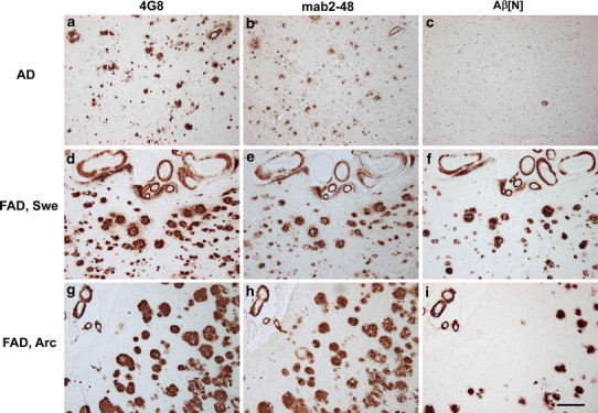
Staining pattern in frontal cortex of sporadic and familial AD brain. Upper panel showing the staining pattern in a sporadic AD case using 4G8 (Aβ 17-24), 2-48 (pGlu Aβ) and Aβ[N] (specific for Aβ at position 1) (a–c). While the staining pattern of 4G8 and 2-48 is comparable, there is no staining with Aβ[N]. Middle panel comparable levels of plaque load with all three antibodies in a FAD case with the Swedish mutation. Note abundant vessel staining with all three antibodies (d–f). Lower panel the same is true for the staining pattern in a case with the arctic mutation; however, Aβ[N] staining is less abundant (g–i). Scale bar 200 μm