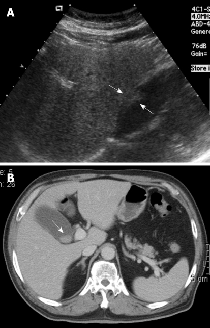
Figure 1.
A 68-year-old woman with intermittent dull right upper quadrant pain for 6 mo. A: Ultrasound (US) showed a sessile polypoid lesion (arrows) with a mildly uneven surface; B: Contrast-enhanced computed tomography (CT) showed an intraluminal polypoid lesion (arrow) with no apparent enlarged regional lymph node.

