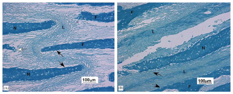
Fig. 4.

Light microscopy of the nasofrontal suture ligament (10 ×). (A) Portion of nasofrontal suture from an older pig tested in tension. The central sutural ligament (L) contains fibers oriented in multiple directions. Black arrows illustrate the fibers oriented to resistant compression and the white arrow fibers oriented to resist tension. Frontal bone (F), nasal bone (N). (B) Tear of the ligament in a nasofrontal suture from a younger pig that underwent compression testing. Black arrows illustrate compression resistant fibers. The central ligament (L) also contains fibers oriented to resist forces from other directions. Although the tension-tested ligament (A) appears stretched and the compression-tested ligament (B) appears compacted, this was not a consistent relationship. Although the bones are thicker in older than younger animals, the trabeculae forming the suture are about the same size.
