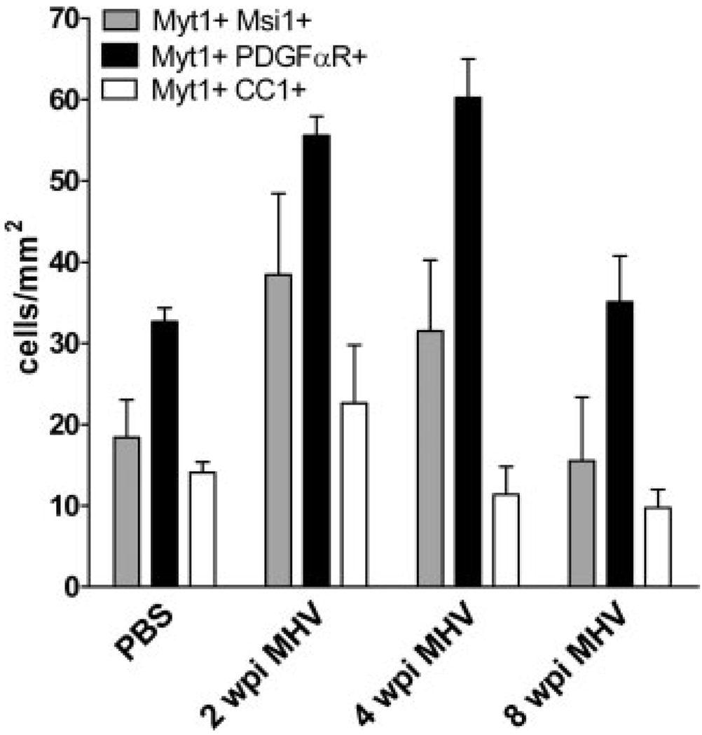
Fig. 4.
Comparison among cell types exhibiting nuclear Myt1 immunoreactivity in PBS control mice and MHV mice throughout disease progression. OP cells immunolabeled with PDGFαR were the principal cell type that expressed Myt1. In addition, the population of Myt1+ PDGFαR+ OP cells increased significantly at 2 wpi (P < 0.001) and 4 wpi (P < 0.001) before returning to PBS control levels at 8 wpi (p > 0.05). Musashi1 (Msi1) neural stem cell marker detected a smaller population of Myt1 expressing cells that was greatest at 2 wpi. Relatively few cells continued to exhibit nuclear Myt1 immunoreactivity after differentiating into mature oligodendrocytes, as indicated by CC1 immunolabeling. PBS, n = 3 mice; MHV, n = 3 mice.

