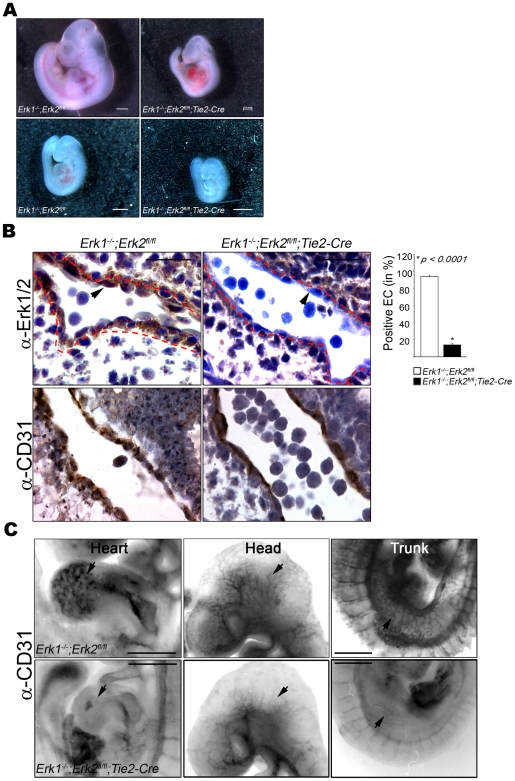
Figure 1. Embryonic Lethality and Defective Angiogenesis in Erk1 − /− ;Erk2fl/fl;Tie2-Cre mutant embryos.
(A) E10.5 day (top) and E9.5 (bottom) embryos; control embryo is at the left, DKO at the right. Bars = 0.5 mm. (B) Consecutive paraffin sections of control (left) and Erk1−/−;Erk2fl/fl;Tie2-Cre (EC-DKO) (right) embryos were stained with anti-ERK1/2 (top) and anti-CD31 (bottom) antibodies. Representative data for E9.5 embryo are shown; a total of 4 embryos were analyzed. Red dotted lines mark the outer lining of the EC layer and arrowheads point to representative EC. Bars = 20 µm. The graphic panel indicates the ratio of ERK positive EC to total CD31-positive cells, expressed as percent positive EC. (C) E9.5 embryos analyzed by whole mount staining with anti-CD31 antibody. Controls are in the top row and EC-DKO mutants in the bottom row. Arrowheads highlight examples of blood vessel staining and branching in the controls that are reduced in the EC-DKO embryos. Bars = 0.5 mm.

