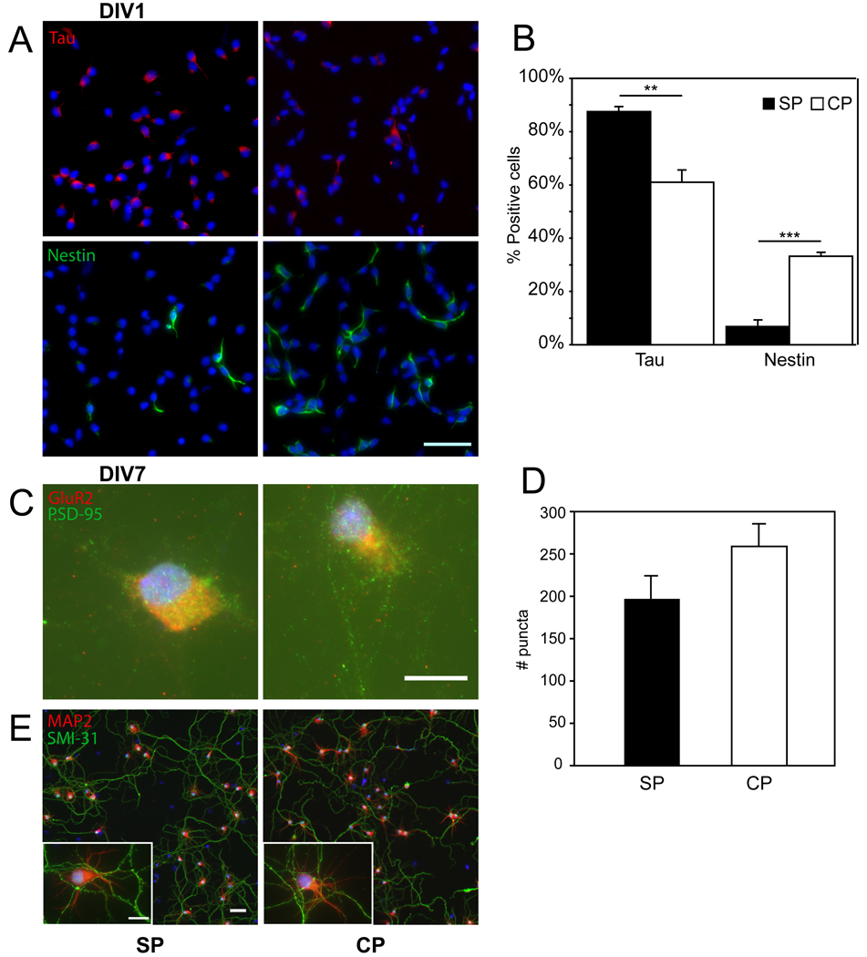
Figure 1.
Immunophenotyping of subplate (SP) and cortical (CP) cells at days in vitro 1 (DIV1) and 7 (DIV7). (A) Tau (red) was used to identify neurons in DIV1 purified subplate cultures (top left) and DIV1 non-purified cortical cultures (top right). Nestin (green) was used to identify neural progenitors in DIV1 purified subplate cultures (bottom left) and DIV1 non-purified cortical cultures (bottom right). Nuclei are counterstained (blue). Scale bar, 50 µm. (B) Quantification of immunostaining from DIV1 (black bars = SP cells, white bars = CP cells, error bars = SEM; ** P < 0.01, ***P < 0.001 by Student’s T-test). N = 4 per culture type. (C) Postsynaptic PSD-95 (green) and presynaptic GluR2 (red) identify puncta in both subplate (left) and cortical plate (right) cultures. Scale bar, 10 µm. (D) Quantification of synaptic puncta from DIV7 (black bars = SP cells, white bars = CP cells, error bars = SEM) (E) Neuronal polarity is shown with dendritic staining of MAP2 (red) and axonal staining of SMI-31 (green) in DIV7 cultures. Scale bar 50 µm, insert, 10 µm.

