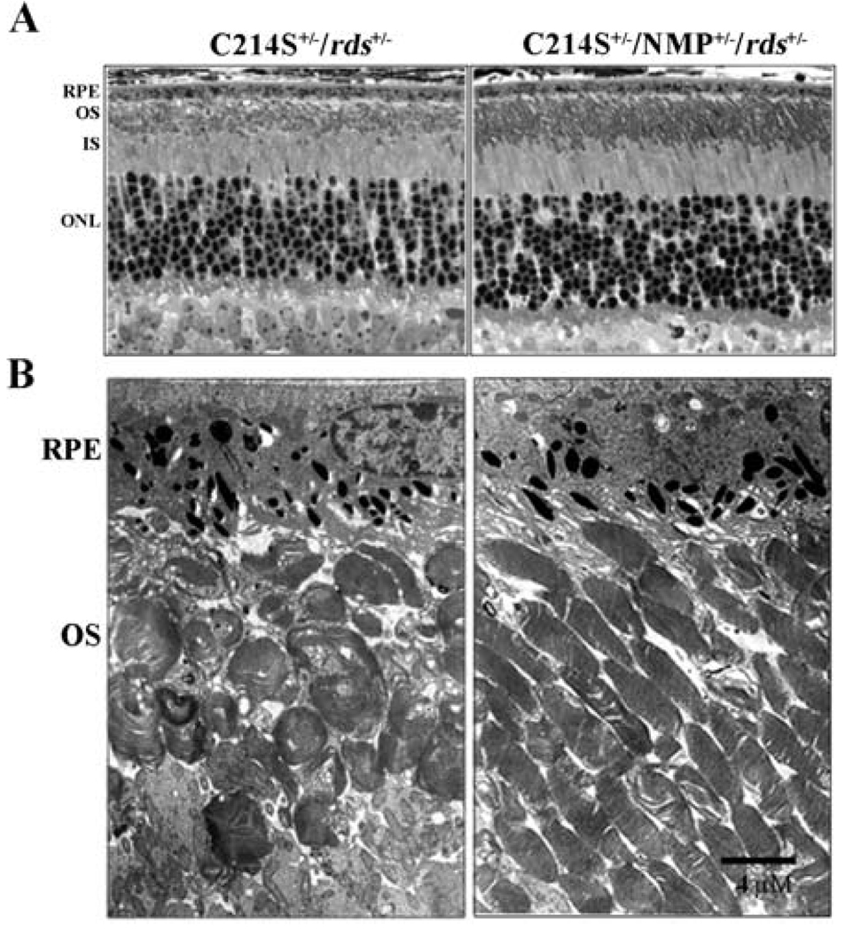
Fig. 3.
Retinal structure in C214S transgenics. (A) Histological evaluation (light microscopy) at 1 month of age reveals an improvement in OS length in C214S+/−/NMP+/−/rds+/− when compared to C214S+/−/rds+/− retinas.(B) Electron microscopy shows amelioration of defects in the alignment and integrity of photoreceptor OSs in double transgenic retinas (C214S+/−/NMP+/−/rds+/−). All tissue sections were evaluated from the superior central region of the retina. Scale bar, 4 µm, (RPE: retinal pigment epithelium; OS: outer segment; IS: inner segment; ONL: outer nuclear layer)

