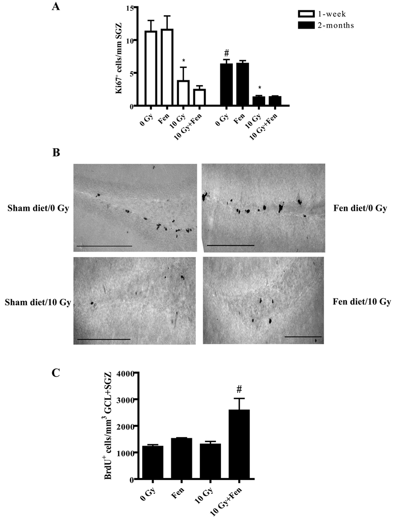
Figure 2. Fenofibrate does not prevent the WBI-induced decrease in proliferation in the SGZ but increases the number of BrdU+ cells in the DG following WBI.
A). The number of Ki67+ cells in the SGZ was significantly decreased at 1-week and 2 months post-WBI; this was unaffected by fenofibrate. Data are presented as Mean ± SEM; n=4 mice/group; an average 6–8 sections were stained using α-Ki67 antibody and counted/animal; * p<0.05 vs. 0 Gy; # p< 0.05 vs. 1-week 0 Gy. B). Representative images showing Ki67+ cells in the SGZ of mice 1 week after sham or WBI with or without fenofibrate administration. Scale bar = 25 µM. C). The number of newly generated (BrdU+) cells significantly increased at 2 months post-WBI in the GCL/SGZ of the fenofibrate-fed WT mice compared to the control diet-fed animals. Data are presented as Mean ± SEM; n=4 mice/group; an average of 6–8 sections were stained using α-BrdU antibody (1:100) and counted/animal; # p<0.05 vs. 10 Gy.

