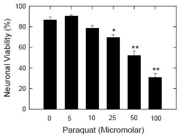
Fig. 2.
Effects of increasing concentrations of PQ on neuronal viability. Primary cortical neurons plated at a density of 300,000 on 35 mm cut-out glass coverslip dishes were exposed to PQ for 24h at a final concentration of 0, 5, 10, 25, 50 and 100 μM. Neurons were labeled with the dyes calcein AM and propidium iodide and the fluorescence images captured on a fluorescence microscope (Nikon Eclipse TE 200). The green (live) and red (dead) cells were counted and data expressed as the percent of total cells in each field that were viable. Data points represent means ± S.E.M. of quadruplicate determinations at each condition in 10 experiments using different cultures. Statistical significance was determined by Student t test (* p<0.01 and ** p<0.001).

