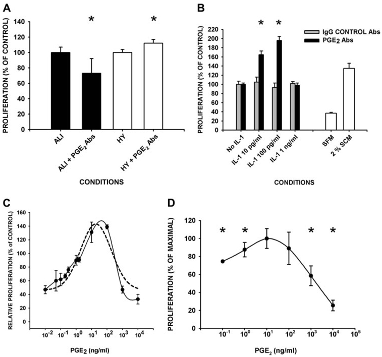
FIGURE 3.
PGE2 mediates proliferation of fibroblasts in pulmonary edema fluid samples, is the downstream effector of low concentrations of IL-1β-induced proliferative suppression, and induces a biphasic proliferative response. A, Serum-starved, human lung fibroblasts (105 cells) were incubated with the indicated edema fluid samples (1/4 diluted in SFM) ± neutralizing PGE2 Ab (10 µg/ml; ALI + PGE2 Abs or HY + PGE2 Abs) or control IgG (10 µg/ml; ALI or HY; n ≥ 3 in quadruplicate/condition). B, Serum-starved, human lung fibroblasts (105 cells) were incubated with the indicated edema fluid samples (1/4 diluted in SFM, A) or the indicated concentrations of IL-1β in 2% SCM for 24 h. The 24-h conditioned medium was harvested and transferred to fresh fibroblast monolayers (1/3 diluted with SFM) ± preincubation with either PGE2 neutralizing Ab (▪) or IgG controls Ab ( ) (at 10 µg/ml; n ≥ 3 in triplicate/condition). C, Biphasic effect of PGE2 on cell proliferation. Fibroblasts were incubated with the indicated concentrations of PGE2 in 2% SCM for 24 h (n ≥ 3 in quadruplicate/condition). Dashed line, mathematical regression; solid line, raw data. D, As in C with a second, independent primary lung cell strain from normal lungs. In A–D, cell proliferation was measured as DNA synthesis by BrdU incorporation. Data are plotted as proliferation relative to the values obtained upon preincubation of the same edema fluid (A) or conditioned medium (B) with control IgG, or with 2% SCM in the absence of PGE2 (B–D). *, Difference between PGE2 Ab and IgG control Ab, or ± added PGE2 at p < 0.05 by t test (A), or ANOVA and post-hoc Student-Newman-Keuls test or Dunnett’s test (B and D).

