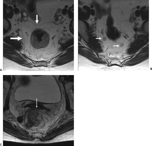
Figure 5.
(A) Magnetic resonance imaging (MRI) scan of a rectal cancer showing the circumferential mesorectal envelope. (B) MRI scan of the rectal cancer patient depicted in (A) showing lymph nodes. (C) MRI scan showing locally advanced rectal cancer that has invaded the posterior wall of the vagina (arrow). (From the Cleveland Clinic Foundation, Cleveland, Ohio. Reprinted with permission.)

