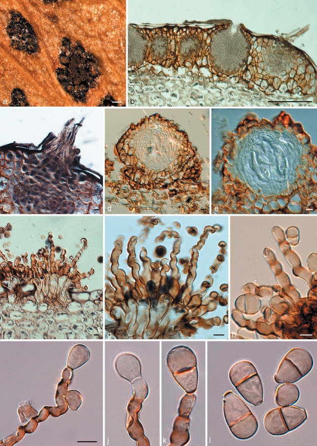
Fig. 3.
Cymadothea trifolii and its anamorph Polythrincium trifolii. a. Ascomata and spermatogonia on the leaf surface; b. vertical section through spermatogonia; c. trichogynes arising from developing ascoma; d, e. vertical section through ascomata; f–h. fasciculate conidiophores arising from leaf tissue. Basal part consisting of tightly aggregated subcylindrical cells that give rise to one or more curved conidiogenous cells with flattened, darkened scars along the length of one side of each conidiogenous cell; i–k. conidiogenous cells with developing conidia; l. mature (0–)1-septate conidia. — Scale bars = 10 μm, except a = 150 μm, b = 120 μm.

