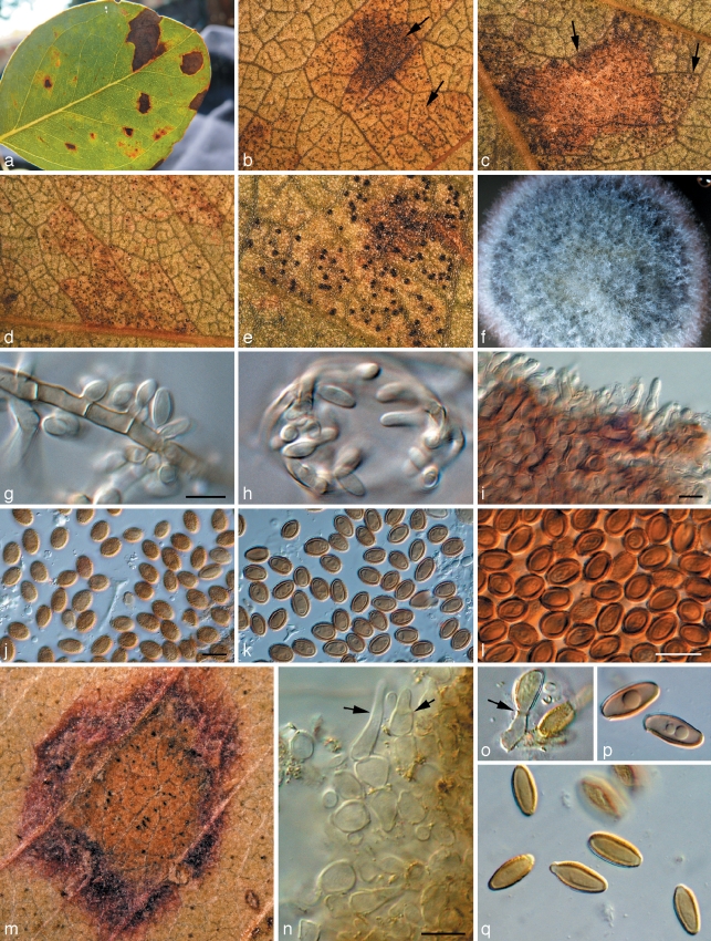
Fig. 4.
Teratosphaeria juvenalis and T. verrucosa. a. Leaf spot with mixed infection of both species; b–e. pycnidia of T. verrucosa on apparently healthy tissue, while those of T. juvenalis occur primarily in leaf spots (arrows). — f–l. Teratosphaeria verrucosa (CBS 113621): colony on PDA; g, h. sporulation on aerial hyphae; i. pycnidial wall with conidiogenous cells in vivo; j–l. conidia in vivo; m–q. Teratosphaeria juvenalis (CBS 110906); m. leaf spot associated with pycnidia; n, o. conidiogenous cells; p, q. conidia. — Scale bars = 10 μm.

