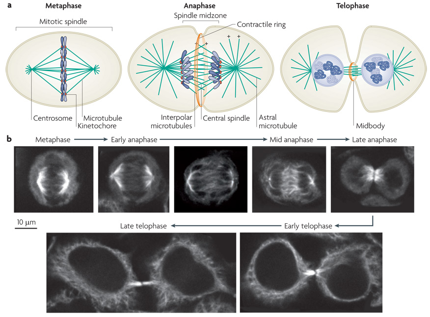
Figure 1. Assembly of the central spindle.
a| Schematic diagrams of the distribution of microtubules and the chromosomes during cell division. In metaphase the chromosomes align on the metaphase plate. At anaphase, the chromosomes move polewards, the central spindle assembles and contractile ring assembly commences. In telophase, after cleavage furrow ingression, the contractile ring compresses the central spindle to form the midbody. Microtubule plus (+) ends are indicated (minus ends, which are positioned at the centrosomes, are not shown). b| Simulated time course of mitotic exit of a cultured human cell line with microtubules labelled by indirect immunofluorescence. At metaphase, the spindle microtubules position the chromosomes on the metaphase plate. In early anaphase, the chromosomes start to move polewards. At mid anaphase, the chromosomes lie at the poles, the spindle has elongated and spindle midzone microtubules are bundled at their overlapping plus ends. In late anaphase, the chromosomes start decondensing and the cleavage furrow has ingressed significantly. In early telophase, the furrow has fully ingressed and the central spindle is compacted into the midbody. In late telophase, the cytoplasmic bridge has narrowed and the cell is prepared for abscission.

