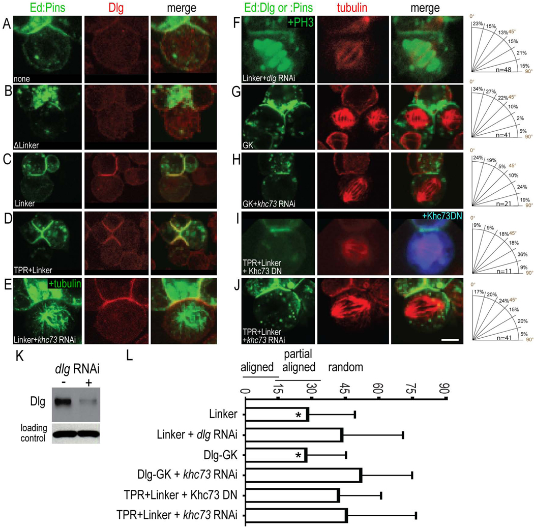
Figure 3. PinsLINKER requires Dlg and Khc73 for spindle orientation.
S2 cells were transfected with Ed:GFP alone or the indicated Ed:Pins domains (green) and immunostained for endogenous Dlg or Tubulin (red). RNAi knockdown was performed for the indicated genes.
(A–E) PinsLINKER recruits Dlg to the cortex.
(F–L) PinsLINKER requires Dlg and Khc-73 for spindle orientation. The mitotic marker PH3 is shown in F. Western blot analysis shows RNAi knockdown of Dlg; detection of α-tubulin demonstrates equal loading of lysates (K). Scale bar 3 µm for A–J.
(L) Quantification of spindle orientation (mean spindle angle ± standard deviation). *, significantly better spindle orientation compared to Ed alone control (p < 0.01); other proteins showed no difference from Ed alone control (p > 0.05).

