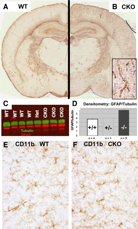
Figure 2.
Cortical GFAP is selectively up-regulated in ERK2 cKO mice. A and B: Coronal sections of 6-week-old wild-type (WT) and cKO mice were processed identically for GFAP immunohistochemistry. A selective increase in GFAP immunoreactivity is apparent in the cortex of a cKO mouse (B) compared with a WT littermate (A), predominantly in perivascular astrocytes (corner inset, B). C: Increased cortical GFAP protein was detected by Western blot in ERK2 cKO mice. Densitometry (D) shows a statistically significant (P < 0.05) increase in GFAP (red) band intensity, normalized to α-tubulin (green) in frontal cortex of cKO mice compared with WT littermates. CD11b immunohistochemistry reveals no evidence of microglial activation in cortical regions showing elevated GFAP (E and F).

