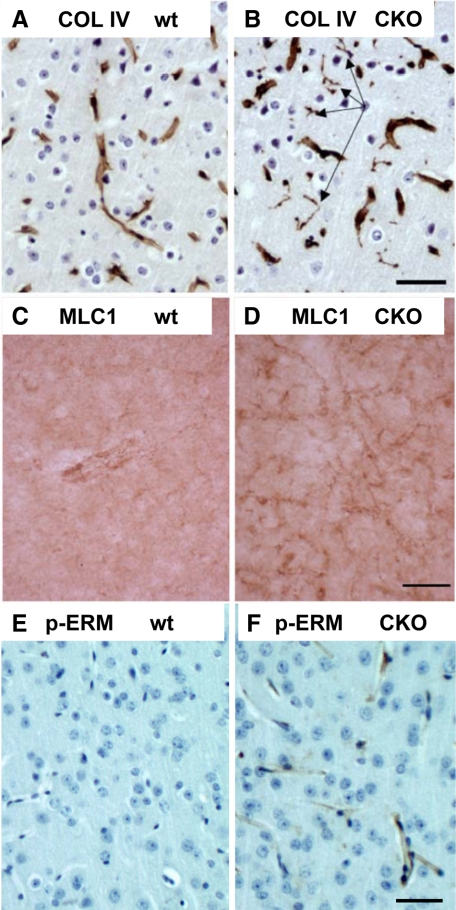
Figure 6.
Evidence for microvascular abnormalities in ERK2 cKO cortex: string vessels, elevated endothelial ERM protein phosphorylation, and elevated MLC1 immunoreactivity. Collagen IV, a nonfibrillar collagen and marker of pericapillary basement membranes, reveals abnormal string vessels in ERK2 cKO cortex (B, arrows) but not controls (A). MLC1, a membrane protein concentrated in distal perivascular astrocyte foot processes, is up-regulated in pericapillary processes in ERK2 cKO (D) compared with wild-type (wt) cortex (C). Phospho-ERM staining, indicating possible growth factor/cytokine activation of endothelium, is up-regulated in cKO cerebral cortical capillary endothelium (F) compared with wild-type (wt) (E). Scale bars represent 50 μm.

