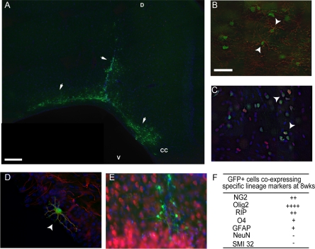
Figure 5.
GFP-OPCs survive transplantation for at least 8 weeks and commit to glial progeny. GFP cells were clearly visible in both gray and white matter at 8 weeks post-transplantation in lesioned animals (arrows, A n = 8, where D: dorsal surface, V: ventral and CC: Corpus callosum). Using confocal imaging GFP-positive (+) cells (green) were identified to express phenotypic markers for NG2-positive cells (arrowhead, red, B), Olig2-positive cells (arrowhead, red, C) and GFAP positive astrocytes (arrowhead, red, D). No NeuN co-labeled cells were observed at 8 weeks (E). Semiquantitative analysis revealed the majority of GFP-positive cells co-labeled with Olig2 and NG2, while none were NeuN co-labeled (F). In all images, Hoechst-positive nuclei were labeled in blue. (−:<1%, +:<25%, ++:25 to 50%, +++:50 to 75%, ++++:>75% of GFP+ cells). Scale bar = 200 μm (A), 100 μm (B, C, E) and 50 μm (D).

