Figure 1.
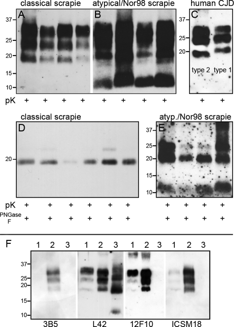
PrPSc typing of sheep scrapie and human sporadic CJD. After treatment with proteinase K alone or combined with PNGase F, Western blot analysis of proteinase K digested classical scrapie cases (A) showed the typical triplet pattern of PrPSc, whereas atypical/Nor98 scrapie samples (B) comprised a multiple band pattern with the characteristic small fragment at 11 to 12 kDa. No differences were detectable in the size of the unglycosylated fragment after additional PNGase F digestion in classical scrapie cases (D). Minor variations of the small fragment of atypical/Nor98 scrapie samples (E) proved to be inconsistent. The differences in molecular size of human PrPSc type 1 (C; right) and type 2 (C; left) were highly reproducible. F: Epitope mapping of ovine scrapie types. Classical scrapie (lane 2) was detectable by antibodies against epitopes in the region of the octarepeats (mAb 3B5) and the helix 1 region (mAb L42, mAb 12F10, and ICSM18), whereas atypical/Nor98 scrapie (lane 3) was detectable by mAb L42 only. Lane 1 is a bovine classical BSE sample.

