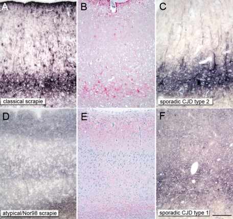
Figure 3.
Obvious parallels in disease-associated PrPSc deposition forms between ovine and human prion types. A reticular/synaptic deposition pattern is observable in the cortices of atypical/Nor98 scrapie sheep (D and E) and sporadic CJD type 1 (F; codon 129 methionine homozygote), while a complex deposition pattern, which is mainly localized in the deep cortical layers, can be seen in classical sheep scrapie (A and B) and sporadic CJD type 2 (C; codon 129 valine homozygote). Ovine tissue: PET blot mAb P4 1:5000 (A and D) and immunohistochemistry mAb P4 1:500 (B and E); human tissue: PET blot 12F10 1:5000 (C and F); scale bar = 300 μm.

