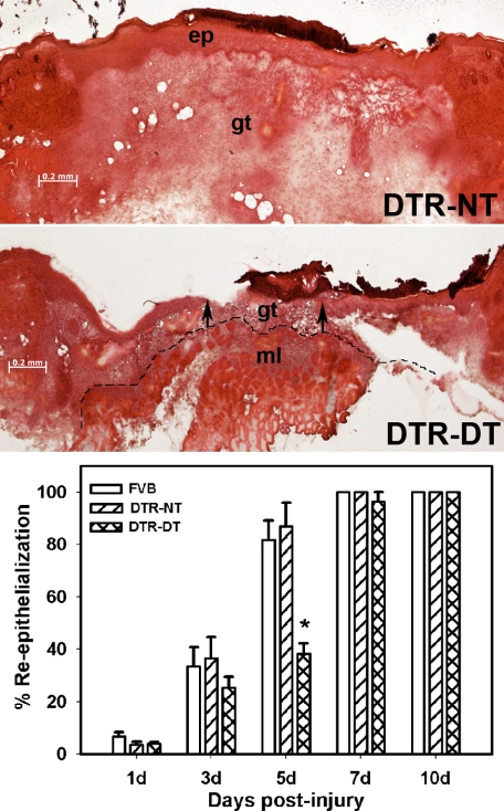
Figure 2.
Macrophage ablation results in delayed re-epithelialization. DTR transgenic mice and FVB wild-type mice were subjected to excisional wounding and either left untreated (NT) or treated with DT. Cryosections of wounds collected on days 1 to 10 postinjury were stained with H&E. Representative sections of wounds on day 5 postinjury showing delayed re-epithelialization in DTR-DT mice (middle) compared with DTR-NT mice (top) (images obtained with a ×5 objective). Arrows indicate ends of the migrating epithelial tongues. ep, epithelium; gt, granulation tissue; ml, subcutaneous muscle layer. Dashed line indicates the border between granulation tissue and subcutaneous muscle layer. Scale bar = 0.2 mm. Bottom: the percentage of re-epithelialization [(distance traversed by epithelium)/(distance between wound edges) × 100] was measured in each section by image analysis. Data are presented as means ± SE; n = 4 to 6 mice/time point. *P < 0.05 when compared to nontreated controls.

