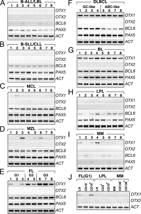
Figure 1.
OTX1 and OTX2 expression in B-cell lymphomas. OTX1 and OTX2 expression analysis in representative pathological samples of B-ALL/LBL (A), B-SLL/CLL (B), MCL (C), MZL (D), FL (E), DLBCL (F), BL (G), LPL (H), and MM (I) shows that OTX2 is not transcribed in any of the pathological samples, whereas OTX1 exhibits high level of expression in all of the patients with FL G3 (E), DLBCL (F), and BL (G). Moderate OTX1 expression is detected in FL G2 (E) and low level of OTX1 is found in most of the FL G1 (E). OTX1 is sporadically expressed at low level in patients with MCL (C), MZL (D), and MM (I); no expression is detected in B-ALL/LBL (A), B-SLL/CLL (B), and LPL (H). High level of PAX5 and BCL6 expression is consistently detected in MCL (C), MZL (D), FL (E), GC like DLBCL (F), and BL (G) samples. J: OTX1 expression in LN, CD19− and CD19+ cells isolated from a representative FL (G1); LN, CD3+ and CD19+ cells isolated from a LPL; and BM, CD138− and CD138+ cells isolated from a MM. G1, G2 and G3 indicate the histological grade of the FL samples. LN tissues are analyzed in all cases, except for lanes 4 to 8 of B where PB tumor cells are used; all lanes of I correspond to BM; lanes 1, 2 of D correspond to splenic tissue and lanes 7, 8 of D correspond to extranodal tissue from gastric biopsies. RT-PCRs are performed at 29 cycles for OTX1, 32 cycles for OTX2, 27 cycles for PAX5 and BCL6, and 23 cycles for β-ACTIN.

