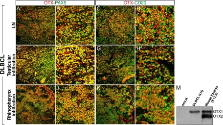
Figure 2.
Immunodetection of OTX1 in DLBCL tissues. Co-immunohistochemistry experiments with OTX and PAX5 (A, B, E, F, I, J) and OTX and CD20 (C, D, G, H, K, L) antibodies in the LN (A–D), testicular (E–H), and rhinopharynx (I–L) infiltrations of a DLBCL show robust expression and full co-localization of OTX1 with both PAX5 and CD20 in tumor cells. M: Western blot analysis showing the OTX1 protein level in extracts from nmLN, DLBCL LN, and mouse embryos at gestational day 12.5 (E12.5). B, F, and J, and D, H, and L are magnifications corresponding to a restricted area of A, E, and I, and C, G, and K, respectively; the mouse and human OTX1 proteins differ by a single amino acid.

