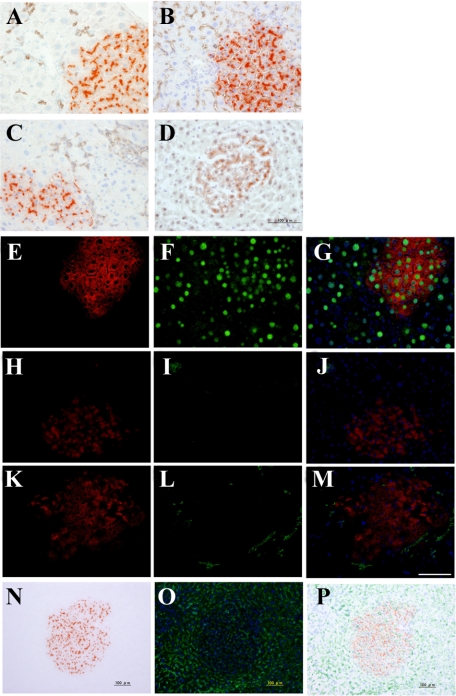
Figure 4.
Characteristics of DPPIV+ foci in the livers after CD44+ cell transplantation. Frozen sections were obtained from livers at 30 days after transplantation of sorted CD44+ cells. Immunohistochemistry and DPPIV-enzyme histochemistry are shown: A combination of DPPIV-enzyme histochemistry and immunohistochemistry for CK19 (A); SE1 (B); CD44 (C); and C/EBPα (D). E–M: Double-fluorescent immunohistochemistry for DPPIV (E, H, K) and C/EBPα (F), CD3 (I), or CK19 (L). N–P: A combination of DPPIV-enzymatic histochemistry (N) and fluorescent immunohistochemistry for SE1 (O). Merged images are combined with DAPI-staining (G, J, M, P). Scale bar = 100 μm.

