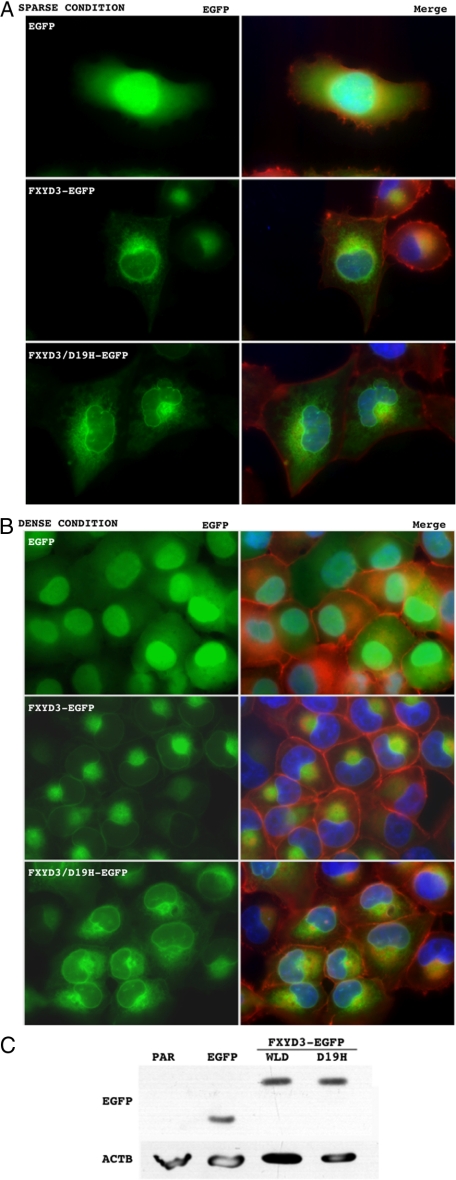
Figure 4.
Subcellular distribution of wild-type FXYD3 and the mutant (FXYD3/D19H), and their effects on morphology in H1299 cells. EGFP (upper panels in both A and B), FXYD3-EGFP (middle panels in both A and B), or FXYD3/D19H-EGFP (lower panels in both A and B) was retrovirally transduced into H1299 cells. After the selection treatment, the surviving cells were harvested and counted, and 1.25 × 10⁴ (sparse condition, A) or 5.0 × 10⁵ (dense condition, B) were re-seeded onto chamber slides (LAB-TEK 2-well chamber, Electron Microscopy Science) and cultured for 24 hours. The cells were fixed with a buffered 4% paraformaldehyde solution and stained with phalloidin-rhodamine (0.1 μg/ml, Sigma) and 4′,6-diamino-2-phenylindole (1.0 μg/ml, Sigma). Green, red, and blue fluorescence indicate the distribution of FXYD3-EGFP (or EGFP alone), filamentous actin, and nuclei, respectively. Left panels in both A and B show representative images of the green fluorescence alone (EGFP). Right panels in both A and B show images layered with green, red and blue fluorescence (MERGE). Expression of the transduced EGFP, the wild-type FXYD3-EGFP (WLD) and the mutant FXYD3/D19H-EGFP (D19H) was confirmed by Western blotting using antibody against EGFP (C, upper panel). Equal loading of protein samples was confirmed by Western blotting using antibody against β-actin (ACTB) (C, lower panel). Par, parental H1299 cells.

