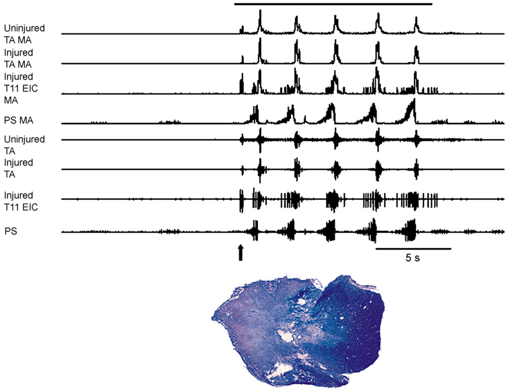
Fig. 2.
Intercostal and abdominal EMGs recorded on the injured and uninjured sides in an anesthetized cat during repetitive cough 5 months following spinal cord hemisection at T9/T10. Arrow indicates the occurrence of an expiration reflex near the onset of stimulation. Following the expiration reflex, five coughs were produced. Robust activity was present bilaterally in the transversus abdominis muscles. The external intercostal EMG on the injured side at T11 was robust. Cough was elicited by mechanical stimulation of the intrathoracic airway with a flexible polyethylene catheter. The solid bar above the EMG records indicates the duration of mechanical stimulation. The hemisection extended across the midline both dorsally and ventrally. TA: transversus abdominis, PS: parasternal muscle, EIC: external intercostal muscle, MA: moving average 50 ms time constant.

