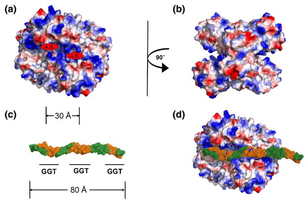
Fig. 6.
Model of the complex between GAPDH and telomeric DNA. (A). Isoelectric-surface representation of a tetramer of GAPDH tetramer, where red is negatively charged, blue is positive and white is uncharged. The active sites are indicated (A.S.). (B). The same representation as (A), but rotated 90°. (C). A model of an 18 nucleotide molecule of ss-telomeric DNA, in CPK format, in which the GGT recognition sequence is colored orange and where nucleotides containing bases that are not required for binding to GAPDH are green. The approximate distance between adjacent DNA-recognition motifs is marked (in Å), which also corresponds to the distance between active sites in the tetramer shown in (A). (D). The same oligonucleotide docked onto the surface of a GAPDH tetramer (similar orientation as (A)) showing the overlap of GGT sequence and active sites.

