
FIG. 4.
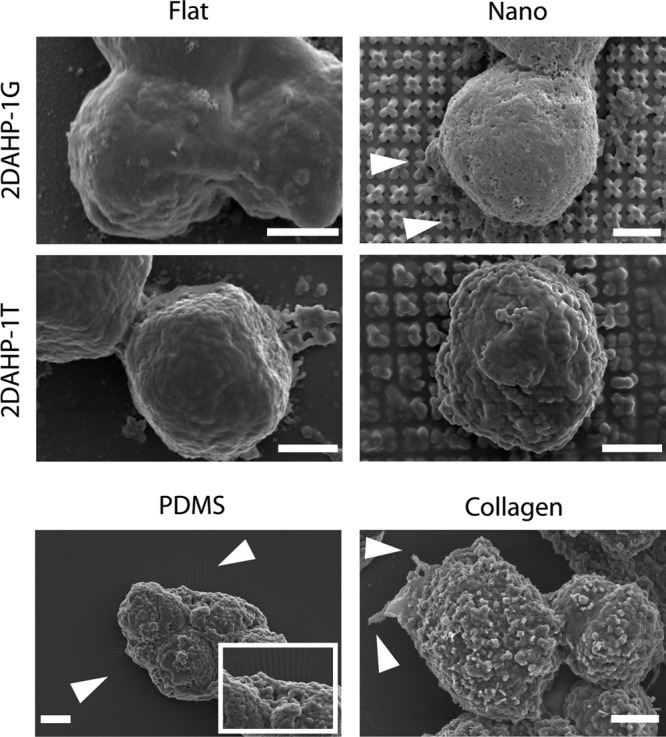
Detailed morphology of cell–substrate interactions. Hepatocytes cultured on all APS substrates exhibited a rounded morphology, while those cultured on PDMS and collagen substrates exhibited a more flattened morphology. Hepatocytes appear to have extended filopodia on nanotopographic 2DAHP-1G, flat 2DAHP-1T, and collagen substrates (see white arrows). Hepatocytes induced wrinkling of PDMS substrates (see white arrows and inset). Again, note the collapsing of the posts on the nanotopographic substrates due to van der Waals forces. Scale bars represent 2 μm.