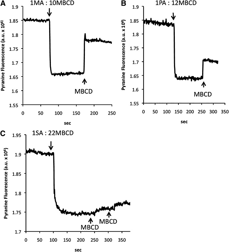
Fig. 2.
Delivery and extraction of MA (A), PA (B), and SA (C) by MBCD to/from lipid vesicles (LUV) measured by pyranine (online fluorescence). Single doses of 4 µM fatty acids complexed with MBCD (as indicated by the arrows; molar ratio FA:MBCD shown) were added to a suspension of LUV containing 0.2 mM entrapped pyranine. MBCD (667 µM) was added to the same LUV suspensions in order to extract the fatty acids delivered to the membrane. LUV was used at a concentration of 400 μM egg-PC in 50 mM HEPES buffer, pH 7.4. One representative experiment is shown in each panel.

