Fig. 3.
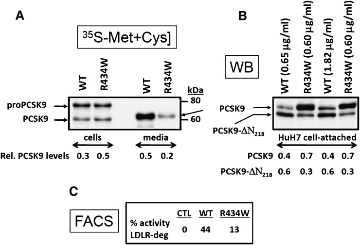
Functional analysis of the PCSK9-R434W variant. (A) HEK293 cells transiently transfected with cDNAs coding for either the V5-tagged PCSK9 (WT) or its R434W variant were pulse labeled with 35S-[Met+Cys] for 4 h and the cell extract and media immunoprecipitated with a V5-mAb and the precipitate separated by 8% SDS-PAGE in 3% Tricine. Autoradiography allowed the identification of the various forms of PCSK9 and image analysis allowed the quantification of the relative amounts of mature PCSK9 in cells and media versus the total levels (17). (B) Spent media from HEK293 cells containing either 0.65 or 1.82 µg/ml of PCSK9 or 0.6 µg/ml of PCSK9-R434W were incubated overnight with naïve HuH7 cells. Following washes, the cells were then detached by EDTA and analyzed by Western blot (WB) using the V5-mAb. Notice that the R434W variant is somewhat resistant to furin cleavage, as evidenced by the lower levels of the PCSK9-ΔN218 form. The relative quantitation of the two forms is presented at the bottom of Figure 3B. (C) The detached HuH7 cells were also analyzed by FACS using a C-7 mAb directed against the LDLR. The levels of the remaining cell-surface LDLR are then expressed as compared with cells incubated with media of HEK293 cells transfected with an empty vector.

