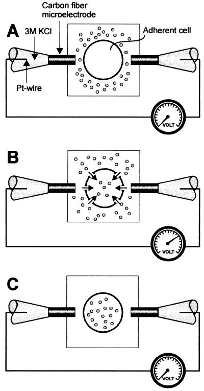
Figure 1.
Schematic picture showing the positioning of the electrodes for single-cell electroporation. (A) Before electroporation. The electrodes are positioned close to the cell with a distance of ≈2 to 5 μm from the cell surface. The buffer contains the solute (dots) to be introduced into the cytosol. (B) Electroporation by application of a rectangular low-voltage pulse. The applied electric field, highly focused over the selected cell, causes membrane-pore formation, allowing the solute in the extracellular solution to freely diffuse into the cell. (C) After electroporation, the pores are resealed, and the solute is trapped inside the cell. After exchange of the extracellular medium, solute molecules are present in the cell but not in the extracellular solution.

