Figure 3.
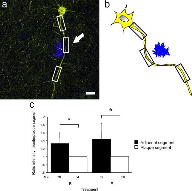
Individual dendritic segments in close proximity to a plaque have decreased neuronal activity. a, b, A neuron and a schematic illustration of a neuron filled with Venus in the vicinity of a plaque (shown in blue). White and black boxes mark the segments where intensity was measured. Note the decreased intensity in the segment close to the plaque indicated by the arrow. c, Dendritic segments farther from the plaque had a statistically significant higher intensity when compared with the dendritic segments nearest to the plaque in unstimulated as well as in stimulated neurons (*p < 0.0001). Scale bar: a, 10 μm.

