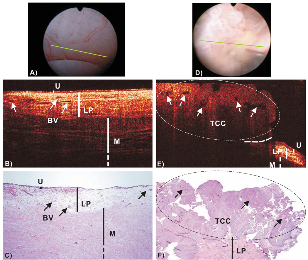
Fig.2.
In vivo surface, cross-sectional COCT and H&E stained histological images of normal human bladder versus a papillary TCC (pT1LG). Image sizes: ~ϕ20mm (A/D) and 4.6mm laterally by 2.1mm axially (B/E, C/F). The morphological details of normal bladder (B), e.g., urothelium (U), lamina propria (LP) and upper muscularis (M) were clearly delineated by OCT based on their backscattering differences; whereas those (e.g., LP, M) underneath papillary TCC (E) diminished. Solid arrows: subsurface blood vessels; dashed arrows: papillary features; dashed circle: TCC (low backscattering), identified by COCT based on increased urothelial heterogeneity; dashed line: boundary with adjacent normal bladder. Diagnoses of the normal bladder: COCT, cystoscopy and histology were all benign, voided cytology was positive. Diagnoses of papillary lesion: COCT, cystoscopy and histology were positive, cytology was benign.

