
Fig. 2.
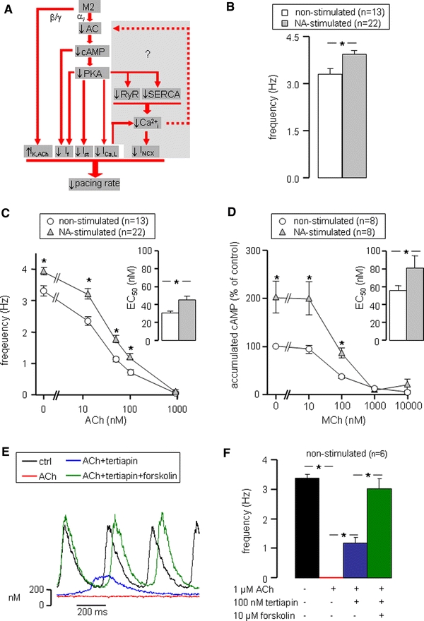
Muscarinic agonist-induced pacemaker slowing. a Muscarinic receptor (M2) stimulation reduces pacemaker rate by down-regulation of inward membrane currents (I f, I st, I Ca,L) through inhibition of cAMP production and up-regulation of the outward I K,ACh. The main objective of the present study is to elucidate the possible role of Ca2+i transients in muscarinic agonist-induced pacemaker slowing (gray area). b Intrinsic pacemaker frequency of non-stimulated (white bars) and 500 nM NA-stimulated (gray bars) SAN cells in the absence of muscarinic agonists. c Frequency–ACh relationship for non-stimulated and NA-stimulated SAN cells. Inset shows average EC50 values of the frequency–ACh relationships of individual cells. d cAMP–MCh relationship for non-stimulated and NA-stimulated SAN cells. Inset shows average EC50 values of the cAMP–MCh relationships of individual cells. e Typical Ca2+i transients of a SAN cell successively exposed to normal Tyrode’s solution (black trace), 1 μM ACh (red trace), 1 μM ACh + 100 nM tertiapin (blue trace) and 1 μM ACh + 100 nM tertiapin + 10 μM forskolin (green trace). f Mean frequency of six non-stimulated SAN cells successively exposed to the solutions of panel E. Quiescent SAN cells (ACh) regained spontaneous activity on inhibition of I K,ACh (ACh + tertiapin), whereas pacemaker frequency was fully restored to control values on additional stimulation of cAMP production (ACh + tertiapin + forskolin). Asterisks indicate significant differences (P < 0.05)