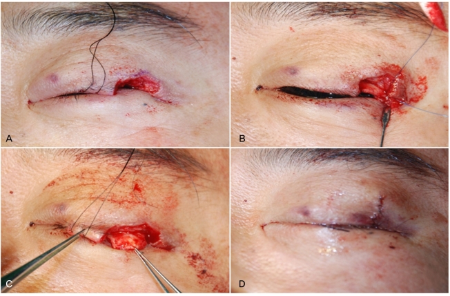
Fig. 5.
Mass excision and reconstruction of the defect. (A) Complete mass excision using a pentagonal shape. Tumor-free margins were confirmed by frozen section biopsy. (B) Reconstruction of the posterior lamella with a tarsoconjunctival sling. (C) Reconstruction of the anterior lamella with a myocutaneous advancement flap. (D) Complete closure of the defect.

