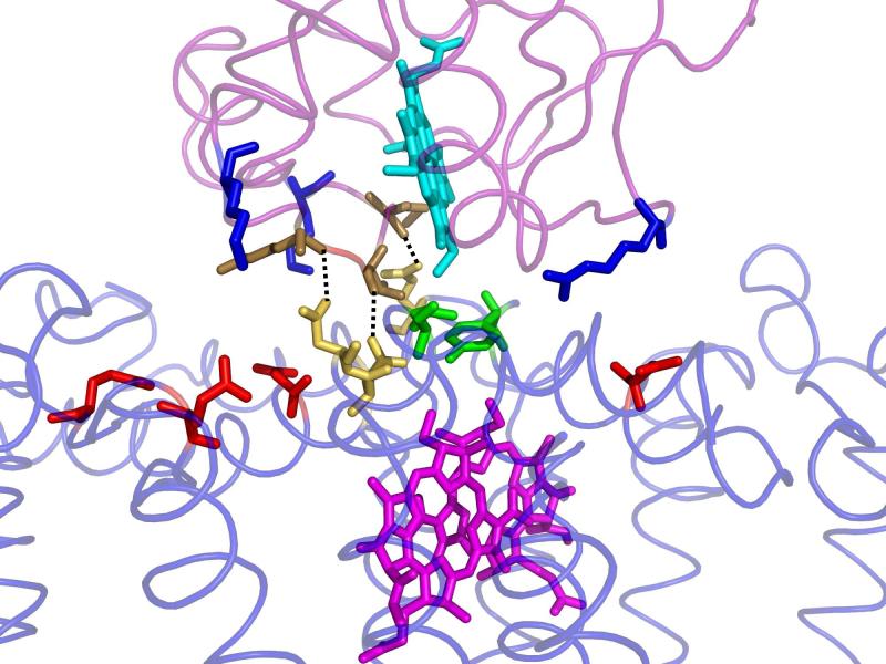
Figure 1.
The structure of the cytochrome c2:RC complex. The cofactors heme (Turquoise) on the cyt and bacteriochlorophyll dimer (Purple) on the RC are connected through a tightly packed interface region containing hydrophobic residues Tyr L162 and Leu M191 (Green) and hydrogen bonding residues Asn M187, Asn M188, and Gln L258(Yellow). Outside this tightly packed central region is a region of solvent separated complementary charged residues; negatively charged (Red) residues on the RC and positively charged (Blue) residues on the cyt (Axelrod et. al (9), PDB 1L9B).

