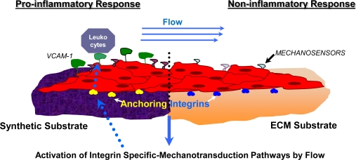
Figure 3.
The effect of sub-endothelial substrate on the activation pattern of pro-inflammatory cellular mechanisms of mechanotransduction. This figure illustrates the biochemical processes induced by fluid shear stress for both a synthetic material and ECM scaffold material. As fluid flows across the EC layer (depicted in red), the mechanosensors at the endothelium surface sense the stress and react by transmitting signals through the transmembrane integrins (illustrated in red and blue). Specific integrin mechanotransduction pathways are thus activated, sending messages from ‘inside-out’ through specific integrins, triggering certain responses. Depending on which type of integrin is activated, various responses occur. Synthetic materials are prone to stimulate a pro-inflammatory response of adhered cell [217] and exposed to the flow, in which presentation on the surface of vascular endothelium of adhesion molecules (for example VCAM-1) induce a subsequent attraction, rolling and adhesion and subsequent attachment of leukocytes to the site.

