
Fig. 4. EGG-4 and EGG-5 inhibit MBK-2 in oocytes and localize to the oocyte cortex.
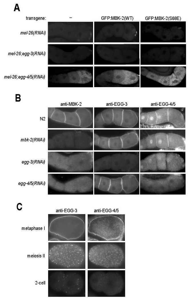
A. Gonads from hermaphrodites with indicated genotypes were fixed and stained with anti-P-MEI-1 antibody. mel-26(RNAi) is used to stabilize P-MEI-1. Note that 1) P-MEI-1 is only seen when EGG-4 and EGG-5 are inactivated by RNAi, and 2) the pattern of P-MEI-1 correlates with oocyte maturation in gonads expressing wild-type MBK-2, and is expanded in gonads expressing MBK-2(S68E). See Sup. Table 1 for numbers and additional genotypes examined.
B. Gonads were stained with anti-MBK-2, anti-EGG-3 and anti-EGG-4/5 antibodies. Note that EGG-4/5 are detected both on the cortex and nuclei of oocytes. The patterns seen after the indicated RNAi treatments suggest that the cortical localization of 1) EGG-3 does not require EGG-4/5 or MBK-2, 2) EGG-4/5 requires EGG-3, but not MBK-2, and 2) MBK-2 requires EGG-3 and EGG-4/5.
C. Embryos co-stained with anti-EGG-3 and anti-EGG-4/5 antibodies. EGG-3 and EGG-4/5 re-localize from the cortex to sub-cortical speckles during the transition from meiosis I to meiosis II. In the 2-cell stage, occasional EGG-3-positive speckles can still be detected. By this stage, the EGG-4 signal is at background levels.