
Fig. 6. EGG-4 inhibits MBK-2 kinase activity in vitro.
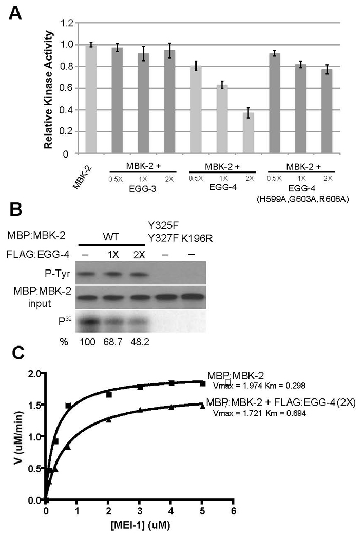
A. 0.5, 1 and 2-fold molar excess of FLAG-tagged EGG-3, EGG-4 and EGG-4(H599A,G603A, R606A) were added to MBP:MBK-2 kinase reactions and the amount of phosphorylated MEI-1 was quantified (as in Fig. 3) after 30 minutes. Levels are expressed as ratio to that observed with MBP:MBK-2 alone (first lane).
B. Same as A, but kinase reactions were split to examine amount of tyrosine-phosphorylated MBK-2 (P-Tyr), amount of MBP:MBK-2 and amount of phosphorylated MEI-1 (P32). Note that addition of EGG-4 lowers the amount of phosphorylated MEI-1 but does not affect tyrosine phosphorylation of MBK-2.
C. Michaelis-Menten plot to compare MBK-2 enzyme kinetics in the presence (2X molar excess) or absence of FLAG:EGG-4.