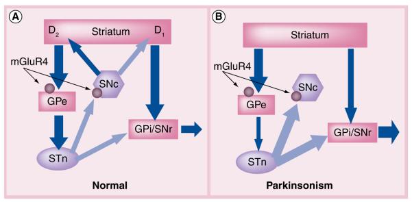
Figure 2. Simplified model of basal ganglia circuitry in normal physiology and Parkinson’s disease.
Excitatory (glutamatergic) projections are shown by light arrows and inhibitory (GABAergic) projections are shown by dark arrows. (A) In the normal BG circuit, direct (striatum to GPi/SNr) and indirect (striatum to GPe) projections to the output nuclei are balanced by striatal dopaminergic tone. D1 receptor-containing neurons stimulate transmission through the direct pathway and D2 receptor-containing neurons inhibit transmission through the indirect pathway. These circuits converge at the GPi/SNr with a balance of inhibitory (via direct pathway) and excitatory (via indirect pathway) inputs to properly regulate inhibitory output to the thalamus. mGluR4 (circle) is localized at the synapse between the striatum and GPe (striatopallidal synapse) as well as synapses between the STN and SNc. (B) In the Parkinson’s BG circuit, the loss of striatal dopamine produces an imbalance in the direct and indirect pathways leading to too much inhibitory tone from the output nuclei. mGluR4-mediated decreases in GABA (striatopallidal synapse) and glutamate (STN–SNc synapse) release are hypothesized to restore balance to the output nuclei and possibly prevent further degeneration of SNc neurons via excitotoxic mechanisms. GPe: External segment of the globus pallidus; GPi: Internal segment of the globus pallidus; SNc: substantia nigra pars compacta; SNr: substantia nigra pars reticulata; STN: subthalamic nucleus.

