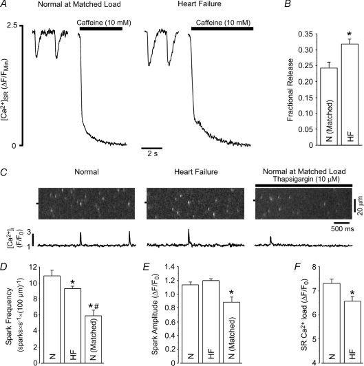
Figure 5. Fractional SR Ca2+ release, Ca2+ spark frequency and Ca2+ spark amplitude are increased in heart failure myocytes.
A, example traces of [Ca2+]SR during 0.5 Hz pacing followed by application of 10 mm caffeine in normal (at matched load) and heart failure myocytes. B, summary data showing the increased fractional SR Ca2+ release during 0.5 Hz pacing in heart failure myocytes when compared to load-matched normal myocytes. Load matching resulted in ΔF/FMin values of 2.60 ± 0.25 (N= 8) and 2.59 ± 0.40 (N= 5) in normal and heart failure myocytes, respectively. C, example images and F/F0 profiles of cytosolic Ca2+ sparks from normal (left), heart failure (middle) and load-matched normal (right) rabbit myocytes. D and E, summary data of Ca2+ spark frequency (D) and Ca2+ spark amplitude (E) from normal (N; n= 37), heart failure (HF; n= 36) and load-matched normal (N matched; n= 17) myocytes. F, SR Ca2+ content was 11% lower in permeabilized heart failure myocytes than in normal myocytes. *P < 0.05 versus normal myocytes, #P < 0.05 versus heart failure myocytes.

