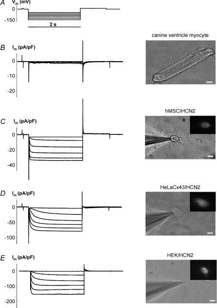
Figure 2. Functional expression of pacemaker current in cells transfected with mHCN2 gene.
Im elicited by hyperpolarizing pulses (A) (from −40 mV to −140 mV) in non-transfected canine ventricle myocyte cell showed little time-dependent activation (B). Im increased with Vm, and hyperpolarization induced voltage- and time-dependent inward currents in hMSC (C), HeLaCx43 cell (D) and HEK293 cell (E) transfected with mHCN2. Fluorescent insets indicate expression of eGFP which is expressed with mHCN2 in these cells (see Methods). 2 mm BaCl2 and 2 mm 4-aminopyridine were added to the bath solution to block voltage-activated K+ currents and unmask HCN2 currents. Scale bar, 10 μm.

