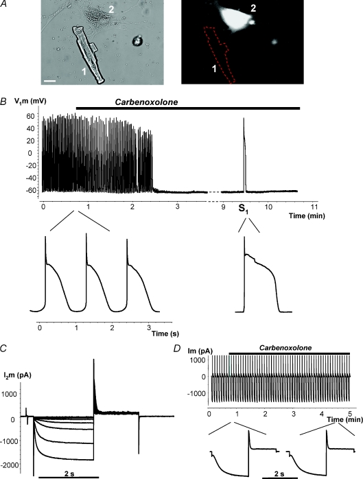
Figure 6. Regulation of spontaneous activity by gap junction blocker carbenoxolone.
A, cell 1, canine ventricle myocyte; cell 2, hMSC transfected with mHCN2. Scale bar, 20 μm. B, spontaneous action potential (AP) recorded from canine myocyte of the above-shown myocyte–hMSC/HCN2 cell pair. Application of 200 μm carbenoxolone abolished spontaneous activity in a time-dependent manner. Application of an external stimulus (S1) evoked a single AP. Inset: APs on expanded time scale before application of carbenoxolone and during application of carbenoxolone evoked by stimulus S1. C, HCN2 currents recorded from hMSC/HCN2 cell after abolishing spontaneous activity with carbenoxolone. D, HCN2 current recorded from HEK293/HCN2 cell, in response to hyperpolarizing voltage pulses to −100 mV. Inset: segments of HCN2 current records on expanded time scale before and during application of carbenoxolone. Application of 200 μm carbenoxolone did not affect HCN2-induced current over time (see details in text).

