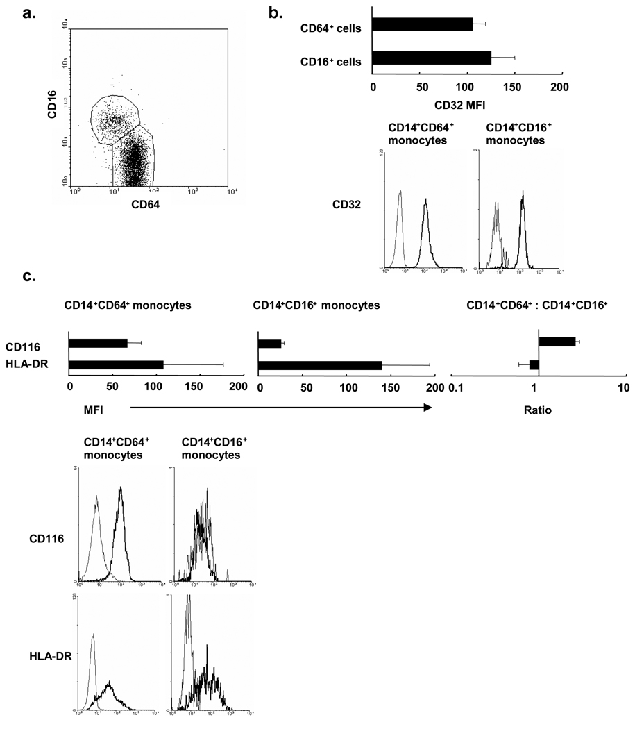
Figure 3. FcγR expression on human peripheral monocyte subsets.
(a) Peripheral human monocytes were labeled with monoclonal antibodies specific for CD14, CD16, and CD64. The dot plot represents CD16 and CD64 expression by CD14 positive monocytes. (b, c) CD14+CD16+ subset and CD14+CD64+ subset were labeled with CD32, HLA-DR or CD116. Data are shown as mean fluorescence intensity (MFI) of three independent experiments +/− s.e.m. (left and center) in addition to representative plots from a single donor.

