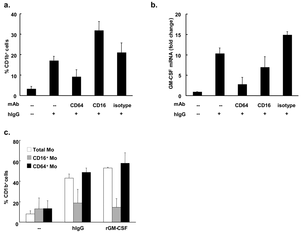
Figure 4. hIgG-triggered differentiation of CD1b+ cells is dependent on CD64.
(a) Peripheral monocytes were stimulated with hIgG (0.2 µg/ml) in the presence or absence of the indicated blocking monoclonal antibodies (10 µg/ml) or isotype control. After two days cells were labeled with anti-CD1b monoclonal antibody and flow cytometry was performed (n=2). (b) Monocytes were stimulated with hIgG (25 µg/ml) in presence or absence of indicated blocking monoclonal antibodies (10 µg/ml) or isotype control. RNA was collected after three hours. Levels of GM-CSF mRNA were measured by qPCR (n=2). (c) Monocytes were separated into CD16+ and CD16− populations using anti-CD16 mAb conjugated magnetic beads. CD16+, CD16− or total monocytes were stimulated with hIgG (25 µg/ml) or recombinant GM-CSF (0.2 U/ml) for two days. The percent of CD1b+ cells was determined using flow cytometry (n=2).

