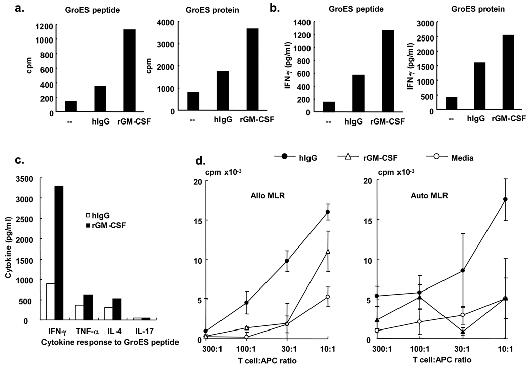
Figure 5. FcγR-differentiated CD1b+ immature dendritic cells activate T cells.
For antigen presentation studies, monocytes were differentiated with either media, hIgG or GM-CSF as above. Cells were cultured with MHC class II-restricted T-cells (1 × 10⁴) and the GroES protein or GroES peptide. We then measured T cell proliferation (a) and cytokine production (b and c). Data are reported from triplicate values and are representative of two independent experiments. (d) For the mixed-lymphocyte reaction, hIgG and rGM-CSF differentiated CD1b+ cells were obtained as above. We obtained T cells from an unmatched donor for the allogeneic MLR or from the identical donor for autologous MLRs using Rosette Sep T cell enrichment cocktail (StemCell Technologies). The T cell:APC ratio varied between 10:1 to 300:1. Proliferation was measured by the thymidine incorporation assay. Data are reported as triplicate values (mean± sem) and are representative of three independent experiments.

