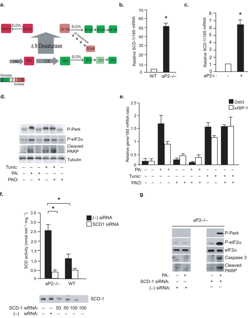
Figure 5. A central role for SCD and C16:1n7 in aP2 mediated lipotoxic signaling.
(A) A summary of the lipid changes that occur as a result of aP2 deficiency in macrophages (LCE and ELOVL; fatty acid elongase for long chain fatty acid). (b)SCD-1 mRNA levels were examined by qRT-PCR in primary peritoneal macrophages at the base line or (c) after treatment of animals aP2-i for 6 weeks (n=6) (data represent mean±SEM; * indicates p<0.05). (d–e) ER stress was induced in macrophages by PA (300 µM) or tunicamycin (2 mM) treatment for 3 hours. Cells were pretreated with PAO (300 µM) PAO, where indicated. P-PERK, P-eIF2–α and cleaved PARP were examined by Western blotting (d) and Ddit3 and sXBP-1 mRNA were examined by qRT-PCR (E). (F) From macrophage lines treated with SCD-1 siRNA (50–100 nM) or scrambled (−) siRNA, SCD activity (upper panel) was examined by an enzymatic assay and SCD protein expression (lower panel) was examined by Western blotting (G) P-PERK, P-eIF2–α and cleaved PARP were examined by Western blotting from lysates of aP2−/− macrophages treated with negative (−) siRNA or SCD-1 specific siRNA (100nM) and treated with or without PA (500 µM) (data represent mean±; SEM; * indicates p<0.05).

