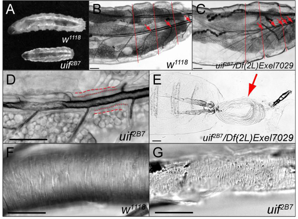
Fig. 5. uif mutant second and third instar larvae have tracheal growth and molting defects.
(A) Brightfield photomicrograph of a 5-day-old w1118 3rd instar larva and a 5-day-old uif2B7 3rd instar larva. Note the smaller overall size of the uif2B7 mutant larva. (B, C) Brightfield photomicrograph of a w1118 2nd instar larva and a similarly sized uif2B7/Df(2L)Exel7029 3rd instar larva showing a tracheal growth defect in the uif mutant larva. Red lines indicate the relative boundaries of the body segments, whereas red arrows indicate the junction of the transverse connective (TC) branch along the dorsal trunk in each metamere. Note that in the wild type larva one TC emanates from each dorsal trunk in each body segment, whereas in the uif mutant larva five TCs emanate from the dorsal trunk in the final two body segments. Also note that the uif mutant larva has less fat than the w1118 larva. (D) uif mutant larvae frequently fail to molt their tracheal cuticle. In this 5-day-old uif2B7 mutant 2nd instar larva the dashed lines indicate the basal membrane of the tracheal cells, whereas the dotted lines indicate the apical surface of the tracheal cells. Note the presence of two layers of cuticle with only the inner or 1st instar lumen being inflated. (E) uif2B7/Df(2L)Exel7029 mutant 3rd instar larva that has failed to complete its molt. The previous epidermal cuticle (arrow) is connected at the posterior spiracles. (F, G) DIC photomicrographs of w1118 and uif2B7 3rd instar larval posterior dorsal tracheal trunks. Note the regular pattern of the taenidial folds in the wild type trachea. Although the trachea in G is uninflated, taenidia are present and are organized into roughly parallel rows that run perpendicular to the long axis of the trachea. Scale bars = 100µm in B, C and E; 50µm in D, F and G.

