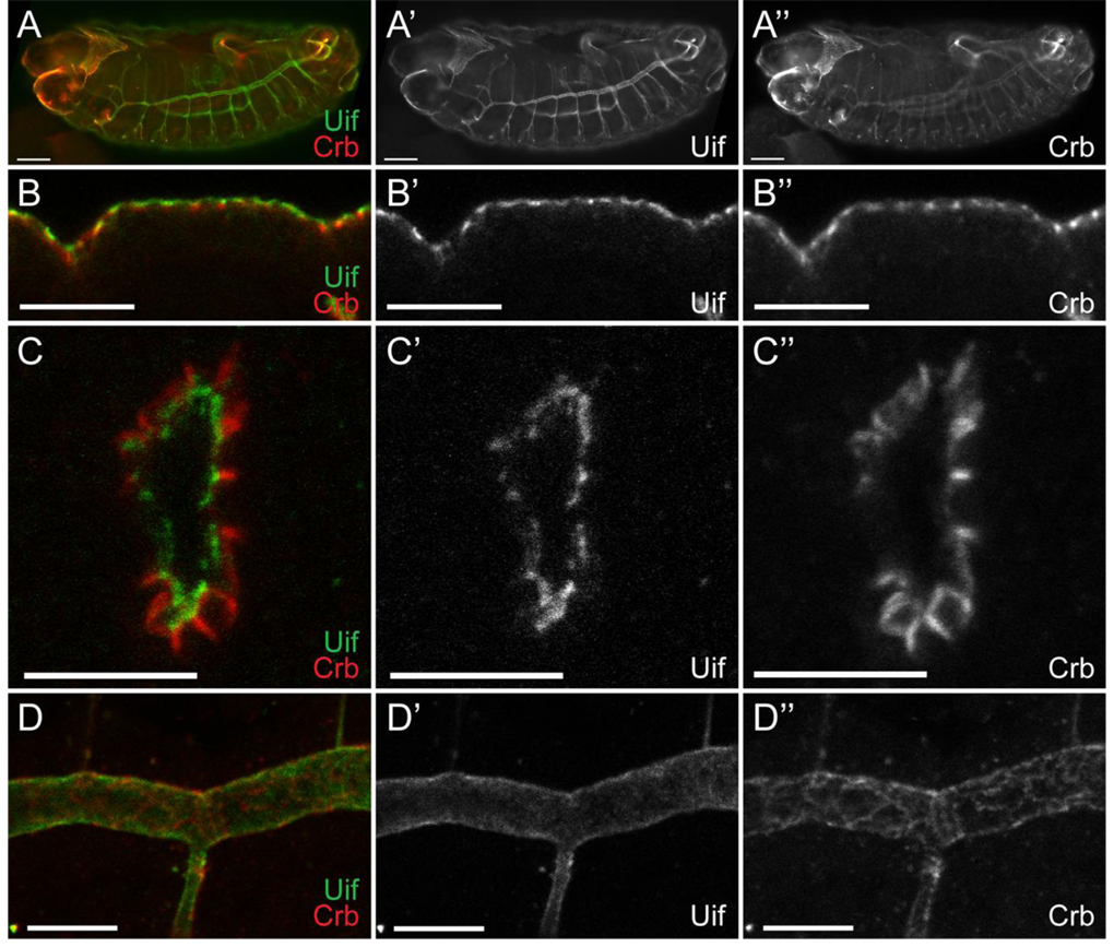
Fig. 6. Uif is expressed on the apical plasma membrane of ectodermally derived epithelial cells during embryogenesis.
(A) Open pinhole confocal micrograph of a stage 15 w1118 embryo double labeled with anti-Uif (green) and anti-Crb (red) antibodies. Individual channels are shown for Uif (A’) and Crb (A”). Uif expression can be detected in the fore- and hindgut, the epidermis and most strongly in the tracheal system. (B–D) Higher magnification confocal optical sections of w1118 embryos stained with anti-Uif (green) and anti-Crb (red) antibodies. Individual channels are shown for Uif (‘) and Crb (”). Tissues include the epidermis at stage 14 (B-B”), the tracheal placode at stage 11 (C-C”), and the tracheal dorsal trunk at stage 14 (D-D”). Note that Uif is expressed in a domain that is more apical than Crbs in all of these tissues. In all cases anterior is to the left. In panel B apical is up. In panel C apical is to the center of the placode. Scale bars = 50µm in A; 10µm in B–D.

