
Figure 5. β Cell Polarity Is Altered in Par1b-Deficient Mice, but Not in PI3K Pathway Mutants.
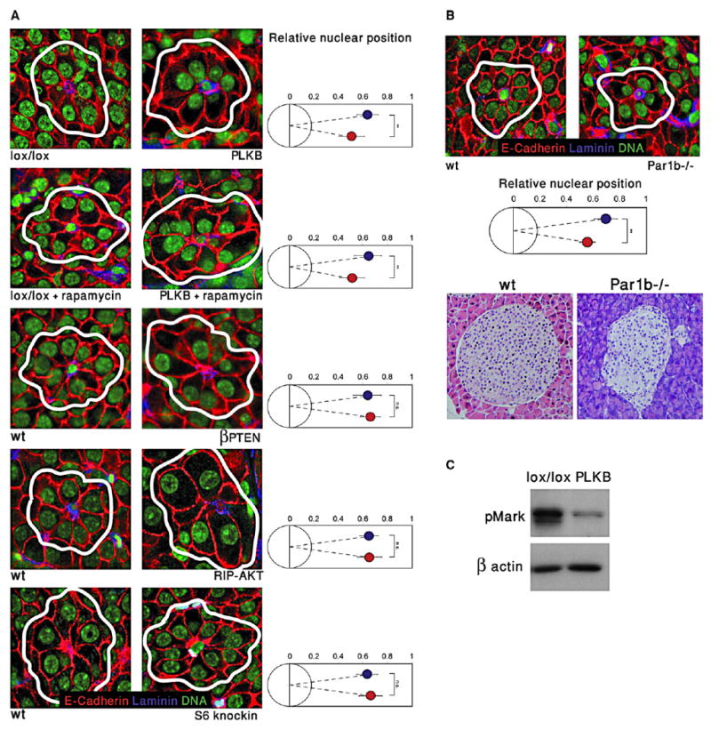
(A) Images of islets from several mutant strains, highlighting rosettes of β cells surrounding capillaries. The position of the nucleus in β cells (right panels) was determined relative to the center of rosettes. Red, E-Cadherin; blue, Laminin; green, nuclei. White lines denote rosettes. Blue, wild-type; red, mutant or transgenic. Original magnification, 600×. Analysis was performed on the same mice as in Figure 4.
(B) Nuclei are shifted to the center of rosettes in Par1b−/− islets. Top, immunofluorescent image (magnification 600×) of representative rosettes from wild-type and Par1b−/− islets, and quantification. Bottom, hematoxylin and eosin staining (400× magnification) showing abnormal distribution of nuclei in Par1b mutant islets.
(C) Reduced phosphorylation of Mark proteins in lysates from PLKB islets, compared with control LKB1lox/lox islets. The upper band represents phosphorylated Mark3, lower band represents Mark2/Par1b. Error bars denote standard deviations.